Home
About ISB
Baliga
Group
High
School Interns
Halobacterium
Our Work
Making Cultures
Streaking Plates
Salinity Lab
Halo Growth with Incubator
Halo Growth without Incubator
Harvesting Membranes
Halo
Crystals
Microbiology
Skills Pt. 1
Microbiology
Skills Pt. 2
Our Mentors
 Institute for
Systems Biology
1441 North 34th Street
Seattle, WA 98103-8904
|
|
|
|
|
|
Harvesting Halo Membranes
Protocol:
We grew 40ml cultures of different strains of halo that started with an
OD600 of 0.05. Two of the flasks were covered in black electrical
tape. In one covered flask and one uncovered flask we replaced
the oxygen in the head space with Argon and put a rubber stopper in the
flask. These were the anaerobic flasks. In the other two
flasks we covered the opening with tin foil. These were the
aerobic flasks. The aerobic flasks were put in the incubator at
225rpm and 36 degrees Celsius. The anaerobic flasks were put in
the same incubator but were not shaken. We grew the aerobic
flasks for 48 hours and the anaerobic for 6 days. When the
cultures had grown we took the OD600 of each. In order to harvest
the cells from the cultures we spun down 35ml of each culture in the
centrifuge at 7000rpm for 10 minutes, then poured off the supernatant.
We then resuspended the pellet in water and DNAase by pipetting
up and down slowly until the solution was homogenous. The water
lysed the halo cells and the DNAase made the solution less gelatinous
and easier to work with. Using the OD600 of the cultures we
calculated the amount of solution from each resuspended pellet to
harvest so that we'd have the same number of cells from each culture.
To harvest the membranes we spun down that amount of each pellet
in an ultracentrifuge at 50,000rpm for 10 minutes. Then we
decanted the supernatant and resuspended the pink part of the pellet in
200 microliters of water. We took out the resuspended pellet,
wiped out the white part, put back in the resuspended solution, and
repeated the harvesting membrane process three times. We used a
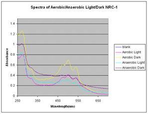
DU800 Spectrophotometer to analyze our membranes using wavelengths from
200nm to 700nm. Then we plotted our data in Excel.
Results:
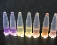
Below is a picture of the membranes of three different halo strains (from left to right: S9, SD23, NRC-1)

Below is the data we collected from out first experiment, as described in the protocol

Next Step:
We repeated this experiment several times using different strains of
Halo (NRC-1, S9, SD23) and different light sources to acheive better
growth and expression of the genes.
|
|
|
|
|
|
|